


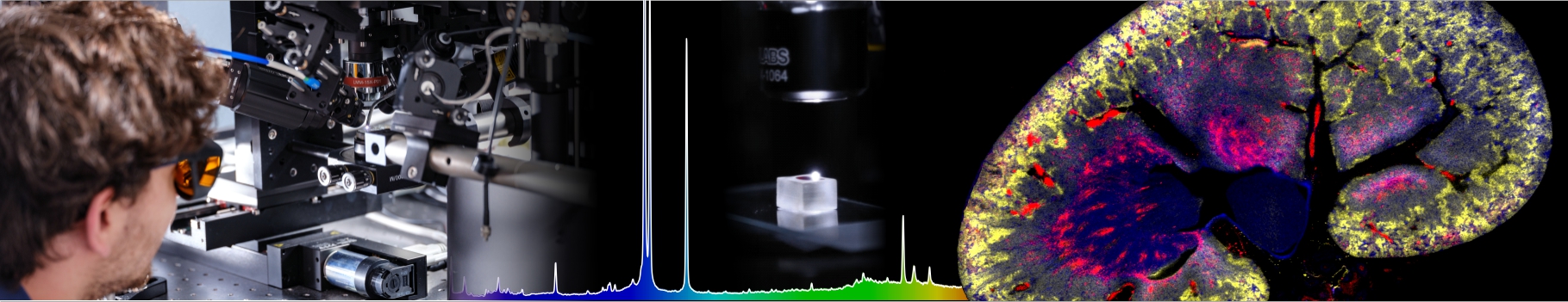

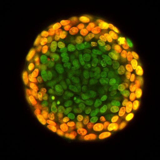
News:New article on : High-speed imaging of mechanical integrity of spheroids for high-throughput pharmaceutical compound screening and pharmacokineticsAlexis Viel, Guqi Yan, Sylvain Monnier and Thomas Dehoux (Biophysics team), in collaboration with colleagues from the LAGEPP in Villeurbanne have published an article entitled « Predicting nanocarriers’ efficacy in 3D models with Brillouin microscopy » in the journal Nanoscale. Today, the evaluation of drug delivery nanosystems in 3D models is mainly explored by means of microscopy techniques that are invasive and require fluorescent labels which modify the composition and fate of the carriers. In recent years, a new quantitative microscopy technique based on Brillouin light scattering (BLS) has been proposed that uses the interaction of laser light with picosecond timescale density fluctuations in the sample. Because it is label-free, all-optical and non-destructive, BLS has gained interest in the pharmaceutical and biomedical fields. In this work, we implemented a fast BLS spectrometer and used the Brillouin frequency shift at the center of spheroids as a quantitative readout for drug efficacy. With a throughput of 50 spheroids in less than a minute, we assessed the efficacy of polymeric nanoparticles loaded with a platinum derivative anticancer drug in reducing the growth of colorectal cancer cells. These results demonstrate that BLS probing of spheroids is a promising tool for high-throughput screening of nanocarriers in 3D models.
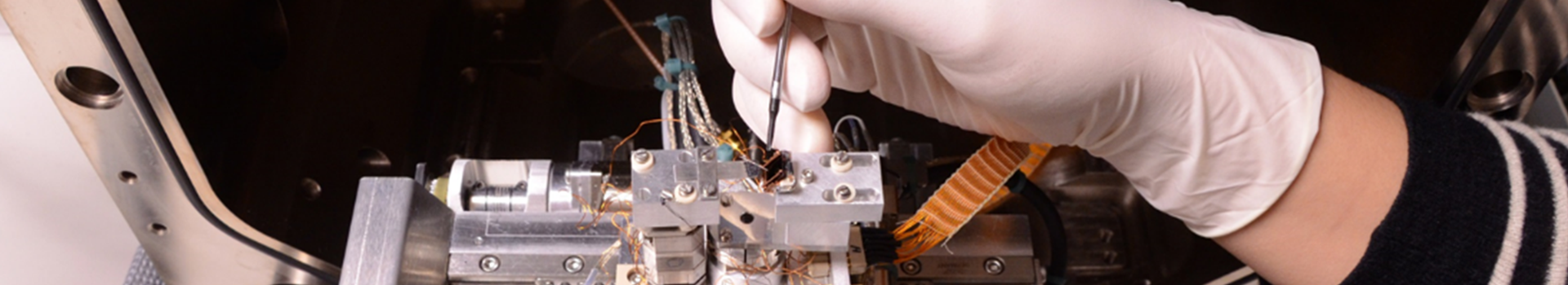
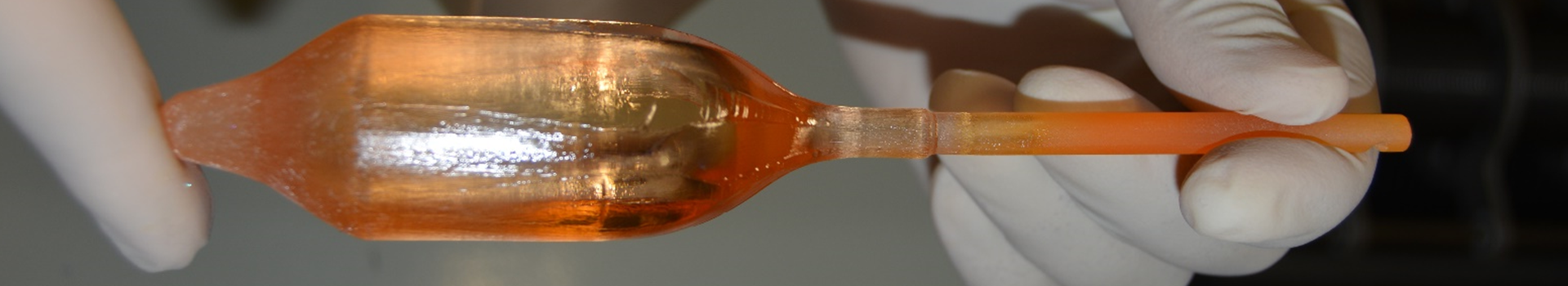
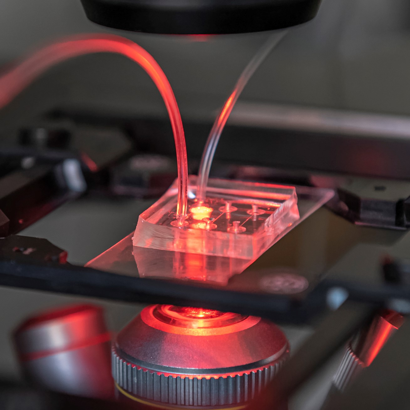
New article on : A platform for parallel electroporation of "mini-tumors"Pauline Bregigeon and Charlotte Rivière (Biophysic team), in collaboration with colleagues from Ecully and Villeurbanne have published an article entitled « Integrated platform for culture, observation, and parallelized electroporation of spheroids » in the journal Lab-On-Chip. The clinical interest in electroporation-based (EPN) therapies has drastically increased in the last few years, as an alternative tumor treatment with reduced side effects and costs. To improve and optimize such treatments, relevant 3D in-vitro cell models are needed, to better reproduce in-vivo context and reduce animal studies. Commonly used in vitro EPN protocols are not adapted to such 3D models. The authors are filling that gap by developping a unique device enabling culture, monitoring, and electroporation of hundreds of regular 3D cell assemblies in parallel, with the same electric field. Such a microsystem could be of primary importance for future studies to better understand the EPN effect in a 3D context and to optimize EPN efficiency and associated drug selection, ahead of in-vivo studies.
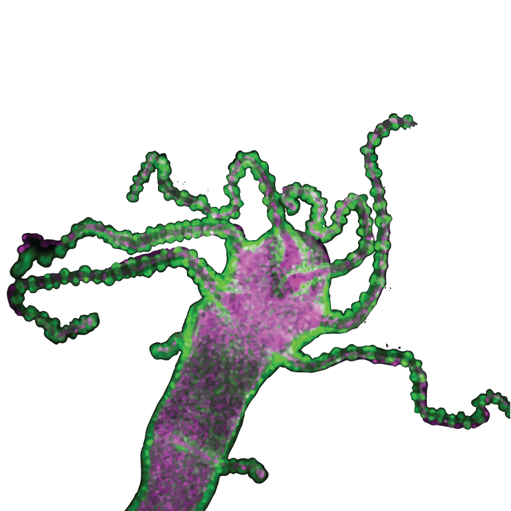
New article on : how oxygen can drive collective migrationOlivier Cochet-Escartin, Christophe Anjard et Jean-Paul Rieu (équipe Biophysique), with colleagues from Lyon, St Etienne and Sendai, published an article entitled "Hypoxia triggers collective aerotactic migration in Dictyostelium discoideum" in the journal eLife.In this paper, the authors demonstrated that the well-known amoeba Dictyostelium is capable of dynamically responding to oxygen gradients created by its own respiration by triggering the collective migration of a dense front of cells, stable over very long time and length scales. These results will impact our understanding of the role of oxygen in the patterning of multicellular organisms as well as in the guidance of cancerous cellular migrations. |
Biophysics TeamThe Biophysics team research topics are centered on the study of cell movements, in particular the emergence of collective behaviors, on the coupling between mechanics and biology for stem cells or tissues (mechano-genetic regulation, tissue rheological properties), and finally on the development of culture and analysis platforms (organ-on-chip) as well as opto-acoustic methods for the characterization of 3D cellular models. These 3D models are multicellular spheroids of cancerous, embryonic or stem cells.
We are developing original methods for measuring forces, mechanical properties and observing the movement and change of shape of cells using homemade routines. Our projects are carried out in collaboration with cancer biologists (we are affiliated with the François Rabelais Institute for multidisciplinary cancer research) and developmental biologists (Hydra, Dictyostelium, plants, protists), engineers of microsystems, polymer chemists, theorists of statistical physics or applied mathematics. |
 |
From cell-cell interactions to emergent collective dynamicsCollective motion of cells is a key biological process that depends on cell migration, communication and interaction with the environment. Using various model systems (Dictyostellium, epithelial monolayers), we study how collective phenomena emerge from the complexity of underlying interactions. |
 |
Mechanics and geneticsThe importance of external mechanical signals on key processes at the molecular scale is increasingly being recognized in the field of mechanobiology. We study these mechano-genetic interactions in the context of cancer, regeneration and development using a combination of techniques from soft matter physics and biology. |
 |
Role of mechanics in cell resistance to therapeuticsWhile the role of mechanics in cancer progression is now recognized, little is still known about its role in cell resistance to cancer treatment. We are addressing this issue by combining different original opto-acoustic and optical imaging techniques. |
 |
Technological developmentsOur team develops tools for quantitative microscopy, image analysis and microfluidics. We have implemented original approaches to study collective motion, the interplay between mechanics and genetics, and the role of mechanics in resistance to cancer therapies. In particular we have expertise in the following techniques.
|