


Methodological developments
4D Traction force microscopy
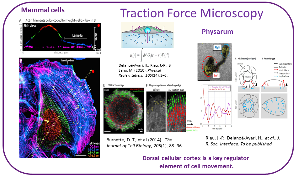
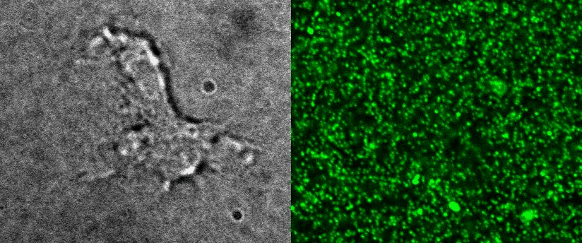
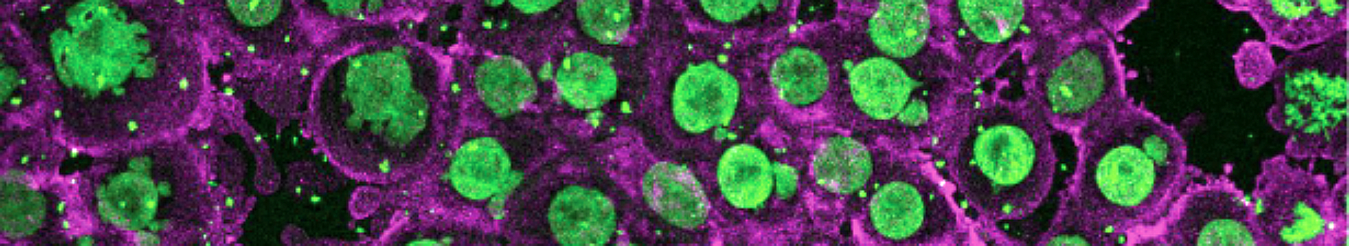
To better understand cell motility in 2D, we have developed measurements of the forces exerted by cells on elastic substrates. For this, we insert fluorescent nanomarkers inside polyacrylamide substrates (whose Young Modulus falls between a few hundred Pascals to a few kiloPascals). When cells moved on the substrate we will monitor the displacement of the markers using 3D timelapse microscopy (See movie). Once the displacement is obtained, the forces exerted by the cells can be calculated back using laws of linear elasticity in semi-infinite mediums.
Optoacoustic sound and light for biology
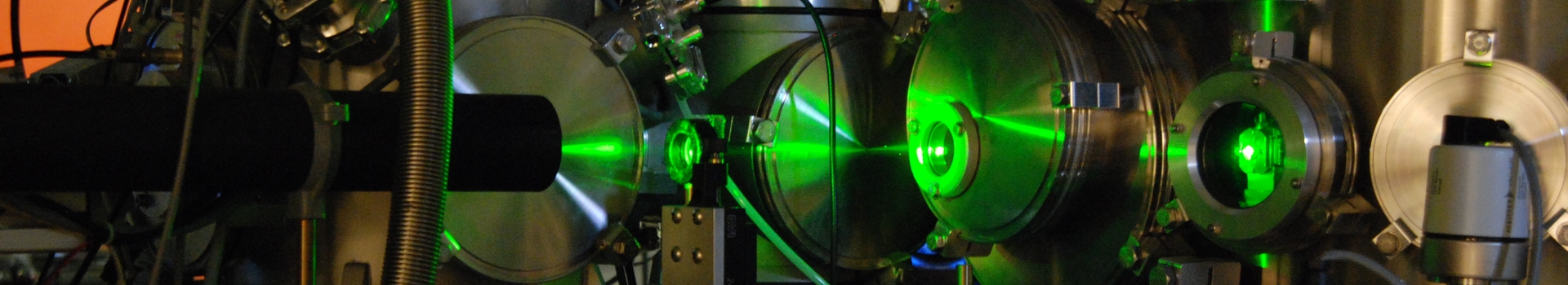
The propagation of ultrasonic waves in biological samples allows probing their mechanical properties, such as rigidity or viscosity. These properties can reveal diseased conditions, but can also shed a new light on the behaviour of cells and tissues. Ultrasound can probe matter at different length scales, from tissue (echography) to single cell (acoustic microscopy), and at different time scales, from millisecond to picosecond (10-12 seconds). Ultrasonic waves are classically generated with piezoelectric transducers. In this project, we produce sound waves using short laser pulses. Such state-of-the-art technology provides innovative means of investigation that are broadband, non-contact, and fully compatible with other advanced light microscopy techniques.
Facilities
Cell culture
Multisite Timelapse microscopy
2-photons microscopy
see NaNoptec website