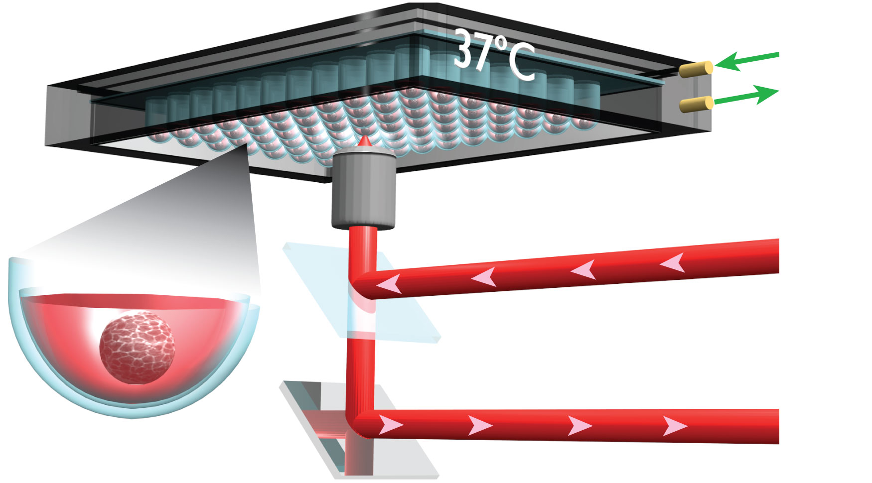
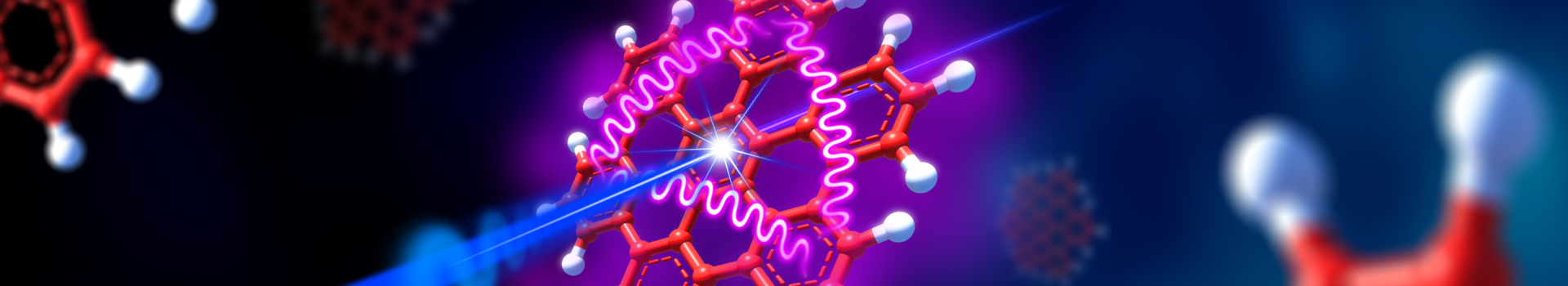

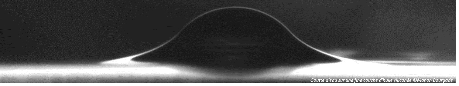
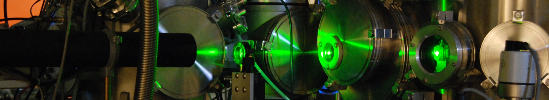

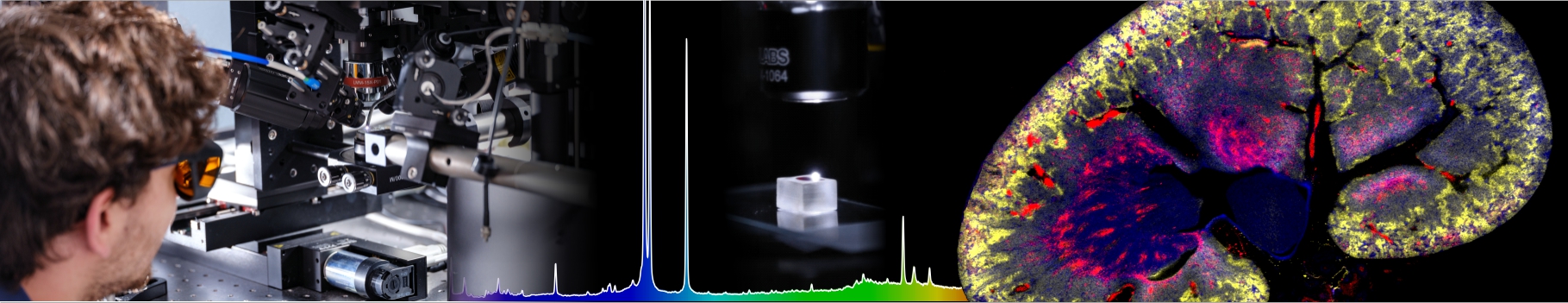
Role of mechanics in cell resistance to therapeuticsThomas Dehoux, Jean-Paul Rieu, Charlotte Rivière We are actively developing systems that recreate in vitro the tumor and recapitulate the main features of its environment. Using 3D-spheroids/organoids models, we are focusing in particular on the effect of mechanics (compression, stiffness, structure of the matrix) on cell resistance to therapeutic agents. Among the therapeutic agents, we are particularly interested in the effect of nano-therapeutics. These nano-objects allow a targeted treatment of cancers, and thus reduce the side effects in the patient. However, their mechanism of action is poorly understood, in particular their penetration and therapeutic efficacy within the tumor. Such effects are difficult to analyze due to the lack of characterization techniques. We have developed 2 original approaches to follow both nanoparticle distribution and therapeutic efficacy within 3D-thick biological samples. For nano-therapeutics, spheroids appear as a good alternative in-vitro models to classical 2D-format Traditional spheroid production and evaluation hampered however to precisely map the distribution of those nano-therapeutics within 3D tissue, and their early effect. Active collaborations : Research Center for Cancer (CRCL, Lyon), Laboratory of Automatic Control, Chemical and Pharmaceutical Engineering (LAGEPP, Lyon), Ampère Laboratory (ECL, Lyon), University of Angers (LARIS). Fundings : ANR, Région AuRA, Mission Interdisciplinaire du CNRS, Plan Cancer, Institut de Chimie de Lyon, Plascan, IMABIO Optoacoustic imagingUsing lasers to generate and detect high-frequency sound waves, we are able to monitor the propagation of the acoustic perturbation in the tumoral tissues and cells. Using this information, these techniques allow measuring the stiffness and viscosity of the sample with a micrometer resolution. The main advantage of such approach is that they are non-contact and label-free.
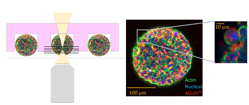
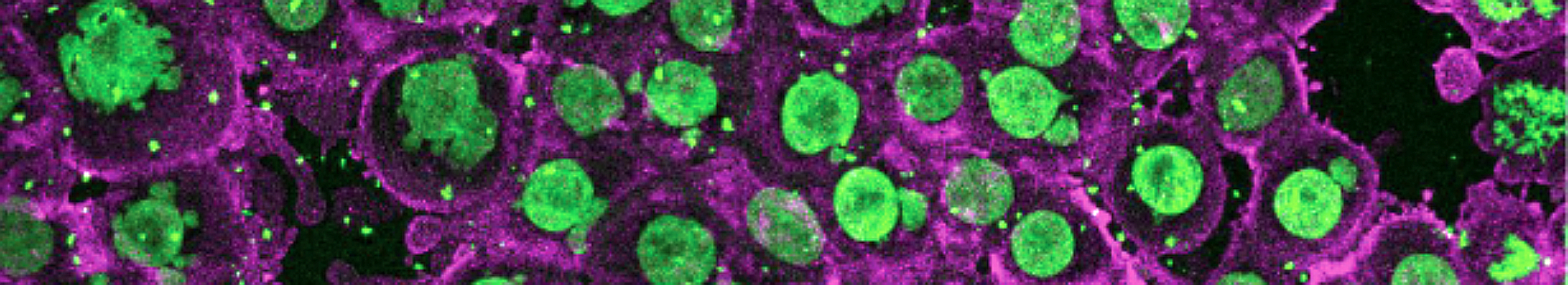
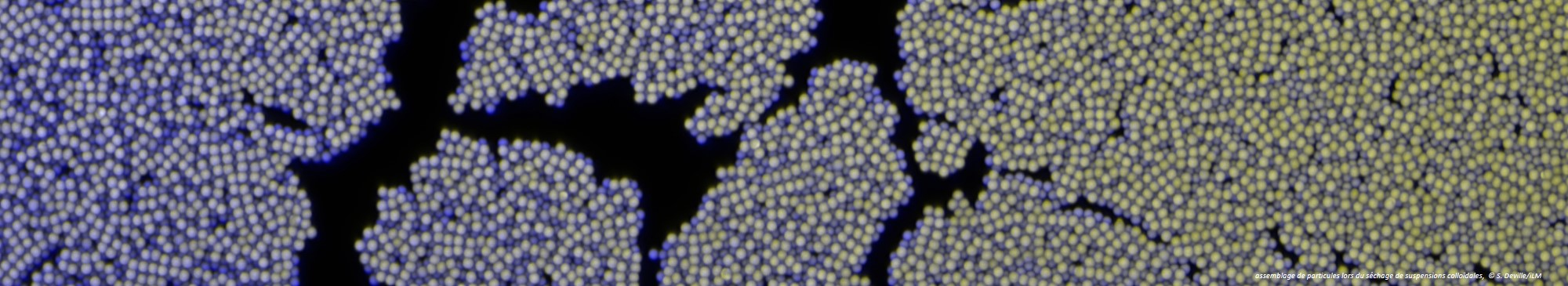
3D optical imaging on whole spheroidsUsing our patented approach described in here, we are currently analyzing the penetration of nanoparticles within colorectal spheroids (See figure). As a proof-of-concept, we are using an experimental therapeutic currently in phase II clinical trials and developed by the FENNEC team of the ILM. Using such approach, we are currently analysing cell response to nano-therapeutics, in order to correlate cell response to nano-therapeutics distribution and localisation within spheroids.
Key publications:Virgone-Carlotta, M. Lemasson, H. C. Mertani, J. J. Diaz, S. Monnier, T. Dehoux, H. Delanoë-Ayari, C. Rivière, and J. P. Rieu (2017), In-depth phenotypic characterization of multicellular tumor spheroids: Effects of 5-Fluorouraciml. PLoS One, vol. 12, no. 11. Rasti, P, R. Huaman, C. Rivière, D. Rousseau (2018), Supervised Machine Learning for 3D Light Microscopy without Manual Annotation: Application to Spheroids, Proceeding in Unconventional Optical Imaging, SPIE, Vol. 1067728, p 80. Lollo, K. Matha, M. Bocchiardo, J. Bejaud, I. Marigo, A. Virgone-Carlotta, T. Dehoux, C. Riviere, J.-P. Rieu, S. Briancon, T. Perrier, O. Meyer, J.-P. Benoit (2018). Drug delivery to tumors using a novel 5FU derivative encapsulated into lipid nanocapsules, Journal of Drug Targeting, 1-26. Margueritat, A. Virgone-Carlotta, S. Monnier, H. Delanoë-Ayari, H. C. Mertani, A. Berthelot, Q. Martinet, X. Dagany, C. Rivière, J.-P. Rieu, T. Dehoux (2019) High-Frequency Mechanical Properties of Tumors Measured by Brillouin Light Scattering. Physical Review Letter, 122, 018101. |